Key takeaways:
- GCA diagnoses made via temporal artery ultrasound were maintained at 1 month and 2 years.
- Ultrasound may speed up diagnostics and reduce need for biopsy and potential resulting complications.
All diagnoses of giant cell arteritis made through temporal artery ultrasound were unchanged at 1 month — and at 2 years, for those with available data — in a study published in Annals of Internal Medicine.
“Unilateral temporal artery biopsy has been recommended as the first-line examination [for potential GCA],” Guillaume Denis, MD, of the department of internal medicine and hematology at Centre Hospitalier Rochefort in France, and colleagues wrote. “However, sensitivity is only about 50% to 70% and specificity depends on many factors such as sample length, pathologist experience, and time elapsed since the start of corticosteroid therapy. In addition, biopsy can lead to complications (pain, hematoma, nerve palsy) and can delay results and mobilization of medical resources.”

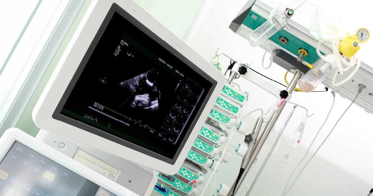
All diagnoses of GCA made through temporal artery ultrasound were unchanged at 1 month — and at 2 years, for those with available data. Image: Adobe Stock
Meanwhile, vascular imaging now plays a role “as important as that” of temporal artery biopsy, Denis and colleagues added. To determine how well temporal artery color Doppler ultrasound “obviates the need” for temporal artery biopsy as a first-line diagnostic test in highly suspected GCA, the researchers conducted a prospective, multicenter study. Participants included 165 patients with suspected GCA referred from general practitioners or ophthalmologists to physicians experienced in GCA at one of six participating hospitals, between August 2016 and February 2020. Follow-up stretched for 2 years.
Patients first underwent temporal artery ultrasound. Those showing bilateral temporal halo sign were diagnosed with GCA and had no biopsy, while those with negative scans underwent biopsy. Patients negative in both were subject to further testing, including full-body CT scan and a multidisciplinary decision-making process.
According to the researchers, 44% (n = 73) of patients were positive on ultrasound. One month later, none demonstrated an alternative diagnosis. Diagnoses remained unchanged at 2 years for all 52 patients with available data.
Among patients with a negative ultrasound, 30% (n = 28) had positive biopsy, and among those with a negative biopsy, 38% (n = 35) were diagnosed based on clinical, imaging and biological data. Overall, 54% (95% CI, 45%-62%) of patients clinically diagnosed with GCA at 1 month demonstrated a positive ultrasound.
“Our study showed that the use of temporal artery ultrasound may be an efficient way to make the diagnosis of GCA in patients with high clinical suspicion and to reduce imaging costs and the need for biopsy, thereby limiting complications and the need for a surgeon,” Denis and colleagues wrote. “In cases of low clinical probability, its role remains to be demonstrated, including in ultrasound-negative cases.”
Sources/Disclosures
Collapse
Disclosures:
Denis reports receiving institutional support for this study from DRCI CHU Poitiers; payment or honoraria from Sanofi Aventis France; meeting and travel support from Leo Pharma, Janssen-Cilag, Novartis Pharma SAS and Takeda France; and participating in a data safety monitoring board or advisory board for Sanofi Aventis France. Please see the study for all other authors’ relevant financial disclosures.