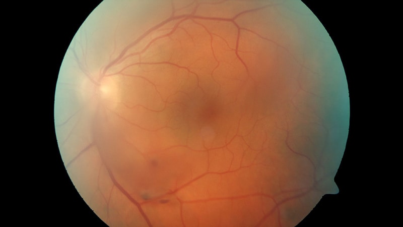
Dilated fundus exams are traditionally used to detect retinal tears in patients with acute posterior vitreous detachment (aPVD). However, alternative techniques like fundus photography and ultrasonography are also effective in identifying the condition. Research presented at the 2024 annual meeting of the Association for Research in Vision and Ophthalmology (ARVO) suggests that these methods have comparable accuracy to the gold standard dilated fundus exams. These findings may lead to easier access to care through telemedicine. Optical coherence tomography (OCT) was found to be accurate in confirming the diagnosis of PVD. Overall, combining fundus photography and B-scan imaging could provide a reliable screening and confirmatory test for retinal tears.
Source link
Imaging Techniques May Match Gold Standard for Retinal Tears
