The human body depends on carefully organized genetic instructions that guide how cells grow and function. Cancer can begin when those instructions become disrupted. Over time, cells may accumulate genetic mistakes that allow them to escape the normal controls that limit growth and division. One of the earliest warning signs in this process is the presence of chromosomal abnormalities, which include changes in chromosome number or structure. These defects can push otherwise healthy cells toward becoming cancerous.
Researchers in the Korbel Group at EMBL Heidelberg have now developed a powerful AI based tool that helps scientists investigate how these chromosomal abnormalities arise. By revealing the conditions that allow these errors to form, the technology may help researchers better understand how cancer begins.
“Chromosomal abnormalities are a main driver for particularly aggressive cancers, and they’re highly linked to patient death, metastasis, recurrence, chemotherapy resistance, and fast tumor onset,” said Jan Korbel, senior scientist at EMBL and senior author of the new paper, published in the journal Nature. “We wanted to understand what determines the likelihood that cells undergo such chromosomal alterations, and what’s the rate at which such abnormalities arise when a still normal cell divides.”
A Century Old Theory About Cancer
The connection between abnormal chromosomes and cancer has been suspected for more than a hundred years. German scientist Theodor Boveri first proposed this idea in the early twentieth century after studying cells under the microscope. His observations led him to suggest that abnormal chromosomal content inside cells could play a role in the development of cancer.
Despite the long standing theory, studying these abnormalities has been difficult. Only a small number of cells display chromosomal defects at any given moment, and many of those cells die (or are killed off) through natural cellular selection. Because of this, researchers traditionally had to search for them manually under a microscope. That process allowed scientists to isolate only a few cells at a time for further study.
Marco Cosenza, Research Scientist in the Korbel Group, began exploring a solution after collaborating with other EMBL teams facing similar technical limitations. Together with colleagues, he helped design an automated platform that integrates microscopy, single cell sequencing, and artificial intelligence. The system is called machine learning-assisted genomics and imaging convergence (MAGIC).
AI Powered “Laser Tag” for Cells
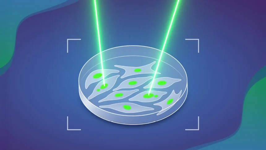
MAGIC functions somewhat like a highly automated version of laser tag. The system scans cells and identifies those that display a specific visible feature. In this study, the researchers focused on a structure known as a ‘micronucleus’.
Micronuclei are small compartments inside cells that contain fragments of DNA separated from the main genome. Cells that contain micronuclei are more likely to develop additional chromosomal abnormalities, increasing their chances of becoming cancerous.
When the system detects cells containing micronuclei, it marks them using a laser. This tagging process relies on a photoconvertible dye, which is a fluorescent molecule that changes the color of light it emits after exposure to light.
“This project combined a lot of my interests in one,” said Cosenza. “It involves genomics, microscopic imaging, and robotic automation. During the COVID-19-related lockdown in 2020, I could really spend some time on learning and applying AI computer vision technologies to the biological image data we had collected before. Afterwards, we designed experiments to validate it and take it further.”
How the MAGIC System Works
The system operates in several automated steps. First, an automated microscope captures a large set of images from a sample of cells. A machine learning algorithm that was trained using manually labeled examples of micronuclei containing cells then analyzes the images.
If the algorithm detects a cell with a micronucleus, it sends the location to the microscope. The microscope then directs a beam of light at that specific cell, permanently tagging it with the photoconvertible dye. Researchers can later isolate these tagged cells from living cell populations using techniques such as flow cytometry. Once isolated, the cells can undergo more detailed study, including analysis of their genomes.
By replacing the slow and labor intensive process of manually searching for micronuclei, MAGIC allows scientists to examine far more cells than was previously possible. In less than a day, the system can analyze close to 100,000 cells.
Discovering How Often Chromosomal Errors Occur
The researchers used MAGIC to study chromosomal abnormalities in cultured cells that were originally derived from normal human cells. Their analysis revealed that slightly more than 10% of cell divisions produce spontaneous chromosomal abnormalities. When the gene p53, a well known tumor suppressor, is mutated, that rate nearly doubles.
The team also examined other factors that may influence the formation of chromosomal abnormalities. These included the presence and position of double stranded DNA breaks within chromosomes.
Broad Potential for Biological Discovery
The research involved collaborations both within and outside EMBL. Key contributors included the Advanced Light Microscopy Facility (ALMF) and the Pepperkok Team at EMBL Heidelberg, Isidro Cortes-Ciriano’s group at EMBL-EBI, and Andreas Kulozik’s team at the German Cancer Research Centre (DKFZ), which is also part of the Molecular Medicine Partnership Unit (MMPU) between EMBL and the University of Heidelberg.
MAGIC is designed to be flexible and adaptable. Although the researchers trained it in this study to detect micronuclei, the underlying AI could be trained to identify many other cellular features.
“As long as you have a feature that can be discriminated visually from a ‘regular’ cell, you can — thanks to AI — train the system to detect it,” said Korbel, “Our system therefore has potential to advance future discoveries in numerous areas of biology.”